Immunohistochemistry Protocol For Frozen Sections

Allow sections to air dry on bench for a few minutes before fixing this helps sections adhere to slides.
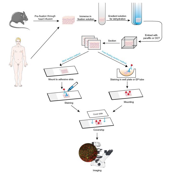
Immunohistochemistry protocol for frozen sections. Fix tissue by perfusing the animal with freshly prepared 4 paraformaldehyde or by immersing it in 4 paraformaldehyde for 4 24 hours at room temperature. Download fluorescent ihc staining of frozen tissue protocol as a pdf. Easy to follow directions describing the step by step experimental procedure.
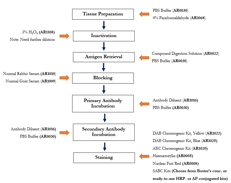
This ihc protocol provides a basic guide for the fixation cryostat sectioning and staining of frozen tissue samples. Frozen tissue indirect method. Frozen section nornally takes less time than paraffin section due to simpler procedures.
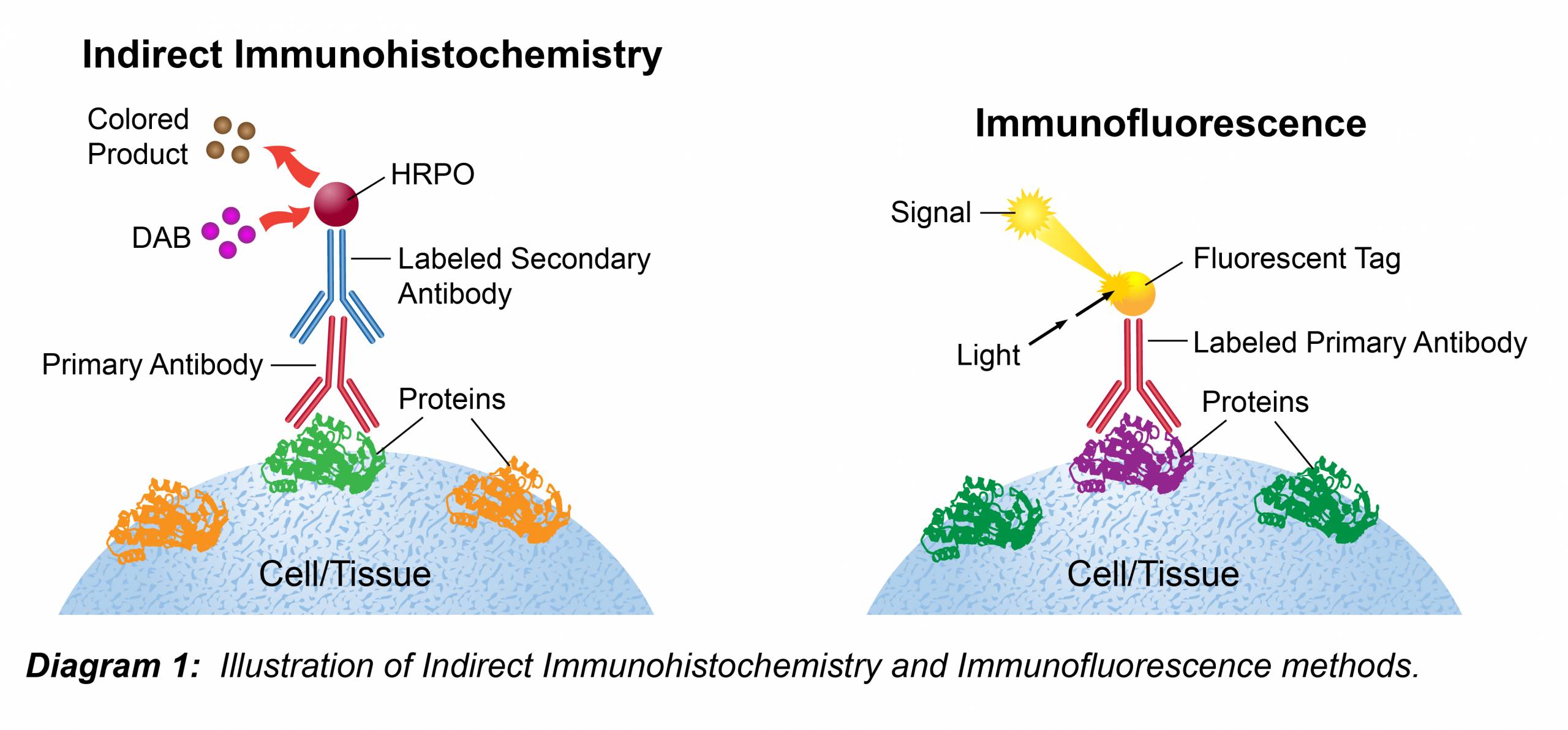
Immunohistochemistry protocol for frozen sections. Uses primary and conjugated secondary antibodies. Section the block at a range of 6 8 µm and place on slides.
Section tissue at a range of 6 8 µm and place on positively charged slides. Immunohistochemistry ihc frozen ihc fr. Some of the steps in this protocol require optimization depending on the sample and antibody being used.
Indirect immunostaining protocol for frozen tissue sections. Fixation temperature and time may. See the next section of this protocol for more information on cryopreservation.
The following immunohistochemistry ihc protocol has been developed and optimized by r d systems ihc icc laboratory for fluorescent ihc experiments using frozen tissue samples. You will also find step by step tips for mouse on mouse staining brdu staining and non antibody based techniques for neuronal staining such as. Frozen tissue direct method.


