Why Are Histological Sections Stained

Collection Of Histological Sections Histologia Microbiologia

The Movat Pentachrome Stain Is For Use In Histological Demonstration Of Collagen Elastin Muscle Mucin Medical Art Microscopic Photography Histology Slides

Histology Art Stained Cells Mad Scientist Science And Nature Medical Science

Pas Staining Human Colon Biology Art Microscopic Photography Histology Slides

Pin On Histology Slides

Staining In Histology Medical Art Science Art Anatomy Art

The tissue is usually sectioned on a cryostat or freezing microtome.
Why are histological sections stained. Because there is no differentiation step background staining can occur especially with charged or treated slides. You observe a tissue that has cells of varying heights. This makes the tissue hard and much easier to cut sections from.
Histopathology refers to the study of tissues that are abnormal or diseased. Histology refers to the study of the individual parts and structures which make up a cell and the relationship between structure and function. The frozen sections are mounted on a glass slide and may be stained to enhance the contrast between different tissues.
Polarity is a property of all normal epithelial tissues. For staining paraffin sections of tissue are normally used. Sections are then stained and examined with the light microscope.
At first glance it appears that the. Pathologists sometimes prefer this type of stain because the non cellular material such as mucin becomes stained with the hematoxylin. This extracellular staining can be an indicator of well differentiated tumors.
Histochemical stains typically haematoxylin and eosin are therefore used to provide contrast to tissue sections making tissue structures more visible and easier to evaluate. Histological sections are stained to enhance the visualization and differentiation of microscopic structures. The role of microvilli is to.
Certain stains change the coloration of cells and tissues significantly different from the color of the original dye complex a phenomenon known as metachromasia. Epithelial tissues are innervated and usually vascularized. Histology stains are used to colour different structures within the cells.
Histology Of The Cervix Showing Clearly Stratified Squamous Epithelium Stratified Squamous Epithelium Squamous Science Geek

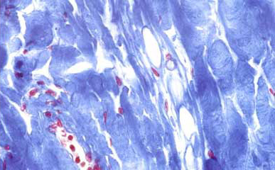
Human Artery Stained With Masson S Trichrome For Collagen Blue And Muscle Red Tissue Types Create Collage Medical Science

This Is A Human Liver Filled With Iron Deposits Blue Gomori S Prussian Blue Stain Tissue Types Blue Stain Human Liver
What Is Histology The Histology Guide
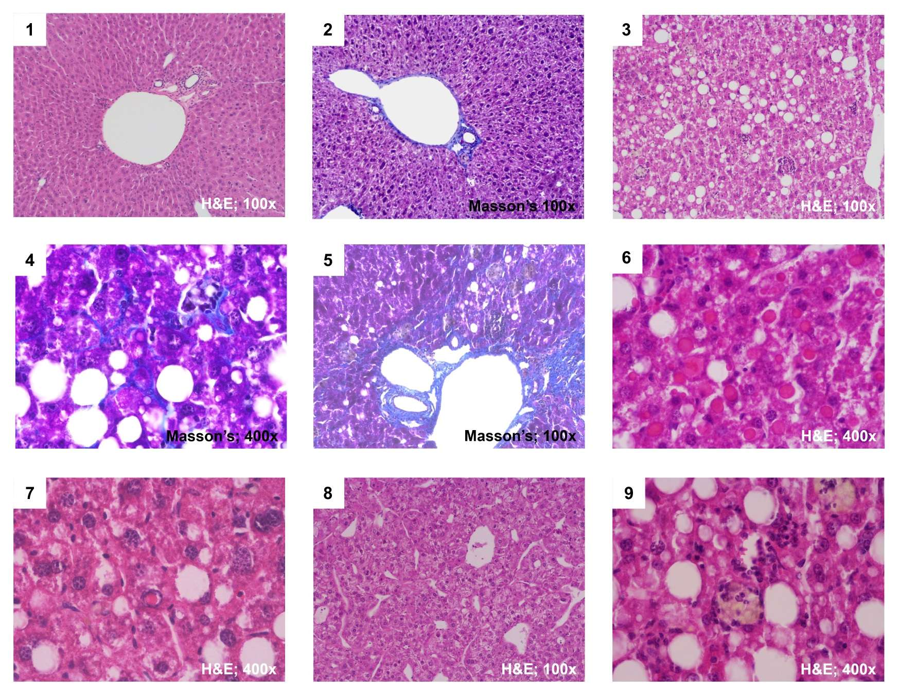
Staining The Liver Ueg United European Gastroenterology

Histology Virtual Slide Box Histology Slides Cells And Tissues School Study Tips
Histological Stains A Brief Overview Of Common Stains

584 Questions With Answers In Histological Staining Scientific Method

Histology Stomach Gastric Gland Pink Cells Parietal Cells Purple Cells Chief Cells Chiropractic Chiropractic Photography In 2020
Histology The John Curtin School Of Medical Research

H E Stain 02 H E Stain Stain Microbiology

Pin On Research Reviews In Biosciences

Pin De Amy Bivens En All Things Histology
